From VOA Learning English, this is Science in the News. I’m Christopher Cruise.
And I’m Faith Lapidus. Today we tell about a World Health Organization report on spinal cord injuries. Then, we tell how American scientists have used stem cells to create lung cells in a laboratory. But first, we report on efforts to create an international database, or electronic record, of brain images. Organizers say the database could help researchers develop better ways to treat chronic, long-lasting pain.
New Brain Image Database Could Help Chronic Pain Sufferers
Many people suffer from chronic pain. Their joints might hurt. They might develop a migraine headache, with sharp pain in the head. Others might have pain in the stomach or abdomen. Many scientists now believe that chronic pain -- in any part of the body -- is linked to the brain. Scientists in California are now developing an international database of brain images of hundreds of chronic pain patients. The goal is to speed up research and develop better ways to treat people whose pain never completely goes away.
Emeran Mayer is a gastroenterologist at UCLA, the University of California Los Angeles. Doctors like him study the stomach and intestines.
“One of the big things in pain research has been the failure to really come up with major breakthroughs in, in treatments.”
Dr. Mayer hopes to develop treatments for people with chronic pain. He is working on a new field that links chronic pain to biological changes in the brain. The Oppenheimer Center for Neurobiology of Stress at UCLA is the headquarters for the first standardized database of brain imaging linked to chronic pain.
“So I think a lot of people now agree with the concept that chronic pain is a brain disease -- it may start anywhere in the body when you have acute pain but once it becomes a chronic pain syndrome it does become a brain disease.”
Kirsten Tillisch is a professor at the Division of Digestive Diseases at UCLA. She says the imaging database lets doctors look at treating the whole body, not just parts of it.
“One of the, the failures of Western medicine and I think our research approaches is how we’ve diced up the human body and how we’ve diced up research into these little silos that work very independently.”
Professor Mayer says Western scientists are starting to look more closely at how the mind, body and environment influence each other.
“Big data, medicine, and analysis is essentially doing the same thing that, you know, the ancient Chinese did by observation. We do it by observing and analyzing very large data sets and trying to see are there patterns in there that, that hang together biologically.”
Professor Tillisch says new technology makes that possible.
“You know when I started this type of work ten years ago there wasn’t -- we didn’t have computing power to do these types of analysis. So, in the brain alone in the, in the last decade it’s really exploded what we could look at.”
The database now has the brain scans of more than 500 patients from North America and Europe. UCLA is working to gather more. Professor Mayer hopes to include brain images of people from Asian countries in the database.
Up To 500,000 Spinal Cord Injuries Per Year Worldwide
The World Health Organization says as many as 500,000 people suffer spinal cord injuries every year. And it says people with such injuries are much more likely to die early.
Recently, the World Health Organization released a report called “International Perspectives on Spinal Cord Injuries.” Alana Officer works at the WHO. She says spinal cord injuries do more than just cause paralysis, or lack of movement.
“There are a lot more associated health problems, such as difficulty with bowel and bladder function, difficulty with sexual function, associated problems around mental health conditions. So it’s much broader than just experiencing paralysis.”
Alana Officer is the WHO’s Coordinator for Disability and Rehabilitation. She says the main causes of spinal cord injuries are traffic accidents, falls and violence. She says some causes are more common in certain areas.
“For example, road traffic crashes are the main contributors of spinal cord injury in Africa and the Western Pacific region. Falls tend to be the leading cause in Southeast Asia and the Middle East. And then we have high rates of violence in certain countries. We have high rates in the U.S. We have high rates in South Africa. And then we’ve also got the non-traumatic causes of spinal cord injuries, such as tumors and cancers, tuberculosis and spinabifida.”
Most people think of tuberculosis as a lung disease. But in some African countries, it is responsible for about one third of the non-violent spinal cord injuries.
The birth defect spinabifida causes damage to the spine. In severe cases, it can affect walking and daily activities. Health officials say they do not yet know the exact cause of spinabifida. But they say it may be linked to genes and the environment.
Alana Officer says more men than women suffer spinal cord injuries.
“There’s a ratio of about two-to-one of males to females. Men tend to be more likely to experience spinal cord injury between the ages of about 20 and 29 -- women, or certainly girls much younger, between sort of 15 and 19. So that’s our first peak in young people. And then we get a second peak, interestingly, in older people. And the major driver of that is falls, tumors, cancer, et cetera.”
She says the main reason people with spinal cord injuries are more likely to die early is lack of medical care.
“A lot of people with spinal cord injuries, certainly in low- and middle- income countries, do not get appropriate emergency response care. Mortality rates are very strongly affected by the quality of the health care system. For example, if you’re in a low-income country, you are three times more likely of dying in (a) hospital following a spinal cord injury than you would be in a high-income country.”
Ms. Officer says many of the causes of spinal cord injury deaths in poor countries are preventable. These include urinary tract infections and pressure sores, also known as bedsores. These are areas of damaged skin caused by a person staying in one position too long. Bedsores are usually not life-threatening problems in wealthy countries.
“People with spinal cord injuries can live pretty much the same amount of time as somebody without a spinal cord injury. There’s a slight difference, but certainly life expectancy has increased considerably in high-income countries. And it’s not the case in low- income countries.”
Experts suggest immediate action if a spinal cord injury is suspected, including immobilization of the spine, restricting its movement. The WHO says that should be followed by what it calls, “care appropriate to the level and severity of the injury, degree of instability of the spine and compression of nerves.” It also suggests “skilled rehabilitation and mental health services.” The WHO notes that up to 30 percent of people with spinal cord injuries show “clinically-significant signs of depression.”
There is currently no cure for paralysis from spinal cord injuries, but many researchers are looking for one. Alana Officer says there is much that can be done to prevent such injuries -- including building safer roads and vehicles, reducing drinking and driving and wearing seatbelts. Other measures include improving safety in sports and the workplace, and adding window guards to windows. She says spinal cord injuries would be reduced if doctors could identify and treat tuberculosis earlier and by improving nutrition to reduce spinabifida cases.
Scientists Create Lung Tissue from Stem Cells
Finally, scientists have used stem cell technology to create working lung cells. Researchers say stem cells also could be used to create new drugs to treat diseases that restrict breathing. And they think the cells could one day create tissue for lung transplant operations. The research is another step toward what is being called personalized medicine.
Over the past several years, scientists have used stem cells and growth factors to force the body’s master cells to create other cells. This process has created heart, intestinal, liver, nerve and insulin-producing cells as possible replacements for diseased organs.
 Human embryonic stem cells are examined under a microscope.
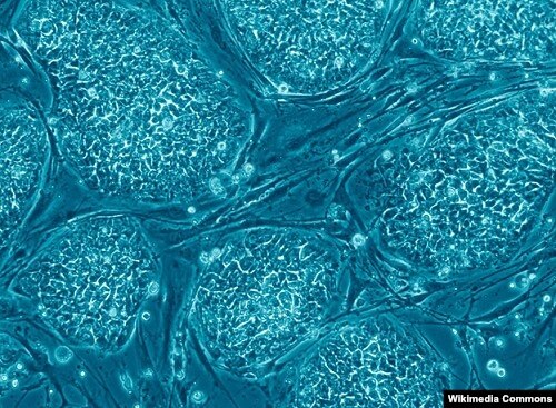
Human embryonic stem cells are examined under a microscope.
Now, scientists at Columbia University in New York City have changed stem cells into working, functioning lung cells, at least in the laboratory. The scientists say this could help researchers develop drugs and transplants to treat deadly diseases.
Hans Snoeck is a professor of microbiology and immunology at Columbia’s medical school. He led the study. He says researchers were able to create six human lung and airway cells. One of the cells produced a substance called surfactant. In the lungs, surfactant keeps the body’s air passageways open, letting people breathe.
“And we know that the cells that we made can make surfactant, can recycle it, can secrete it again. So we know that they do what they’re supposed to do. And these cells are very essential in lung diseases.”
He adds that impaired surfactant production causes pulmonary fibrosis, a deadly lung condition.
In experiments, researchers showed they could make embryonic stem cells become lung tissue. They used proteins that tell the body’s master cells to become lung tissue. Some people have criticized the use of embryonic stem cells because they come from human embryos. That is why the researchers turned to what they call induced pluripotent stem cells. These are skin and other kinds of cells that can act like progenitor cells.
All of the experiments produced human lung tissue. The researchers placed this tissue into the kidneys of mice. Dr. Snoeck says the implanted cells grew and began acting like lung tissue. Researchers will be able to use the cells as a model for treatments for disabling and deadly lung diseases, like cystic fibrosis.
Dr. Snoeck says lung transplants often are not successful. But in the future, he says, scientists may be able to engineer transplantable donor lungs by removing all of their cellular material. They would then seed the remaining structures with lung cells created from the skin cells of the transplant patient.
“In this way we hope to be able to one day -- but this is years away -- to make a lung that a recipient -- someone who is in need of a transplant -- will not reject because all the cells in this lung are made from the same patient.”
A report on the creation of lung tissue from stem cells was published in the journal Nature Biotechnology.
This Science in the News was written by Christopher Cruise. Our producer was June Simms. I’m Faith Lapidus.
And I’m Christopher Cruise. Join us again next week for more news about science on the Voice of America.
Loading lesson...